养猪宝
当前位置: 猪病防治 > 猪传染病 > 正文
中国养猪网 中国养猪网

非洲猪瘟病毒在掩埋野猪尸体中的存活情况(上)

来源: 猪译馆   2020-06-12 14:36:14   查看:  次


译者的话:


已知非洲猪瘟病毒(ASFV)在环境中非常稳定。因此,受感染野猪的尸体可以作为ASFV的主要宿主,从而有助于在野猪群中局部维持和传播该疾病。为了尽量减少这一风险,在ASF疫区清除野猪尸体被认为是有效控制疾病的关键。如果野猪尸体无法清除,通常就地掩埋处理,以避免野猪直接接触感染源。
在本研究中,在立陶宛不同时间和地点掩埋的感染了ASFV的野猪尸体被挖掘出来,并通过体外试验和定量PCR重新检测了传染性ASFV的存在。另外还对可能被体液污染的土壤样本进行了病毒基因组检测。在20个埋葬点中的17个,挖掘出的尸体样本都发现了ASFV基因组然而,在所有的动物尸体样本中,都无法分离出ASFV。在7埋葬点的土壤样本中含有ASF病毒DNA。这些结果出人意料地否定了在野猪尸体中不受埋葬时间影响的传染性ASFV的长期存在。

非洲猪瘟病毒在掩埋野猪尸体中的存活情况(上)


African Swine Fever Virus Survival in Buried Wild Boar Carcasses – Part 1 


作者Authors:


Claire Guinat, DVM, MSc, MRCVS

Andrey Gogin, DVM, PhD 欧洲食品安全局 European Food Safety Authority

Sandra Blome, DVM, Dr med vet

Guenther Keil, DipBiol, DrRerNat

Reiko Pollin, DipBiol, DrRerNat 里德里希·吕弗勒研究所 Friedrich-Loeffler Institut

Dirk U. Pfeiffer, PhD 英国皇家兽医学院兽医流行病学经济学和公共卫生研究组 Royal Veterinary College, Veterinary Epidemiology, Economics and Public Health Group

Linda Dixon, BSc, PhD, 英国皮尔布莱特研究 The Pirbright Institute


摘要Abstract

自从1957年非洲猪瘟(ASF)首次欧洲野猪群中爆发以来,人们一直在讨论死于该病的动物尸体中病毒的存活问题。已知非洲猪瘟病毒(ASFV)在环境中非常稳定。因此,受感染野猪的尸体可以作为ASFV的主要宿主,从而有助于在野猪群中局部维持和传播该疾病。为了尽量减少这一风险,在ASF疫区清除野猪尸体被认为是有效控制疾病的关键。如果野猪尸体无法清除,通常就地掩埋处理,以避免野猪直接接触感染源。在本研究中,在立陶宛不同时间和地点掩埋的感染了ASFV的野猪尸体被挖掘出来,并通过体外试验和定量PCR重新检测了传染性ASFV的存在。另外还对可能被体液污染的土壤样本进行了病毒基因组检测。在20个埋葬点中的17个,挖掘出的尸体样本都发现了ASFV基因组然而,在所有的动物尸体样本中,都无法分离出ASFV。在7埋葬点的土壤样本中含有ASF病毒DNA。这些结果出人意料地否定了在野猪尸体中不受埋葬时间影响的传染性ASFV的长期存在。在这种情况下,必须进一步研究从动物尸体样本中分离出的ASFV的敏感性以及动物的易感性和口服接种所需的剂量。此外,还需要研究考虑在野猪感染循环其他的ASF感染源和驱动因素。

Since the first introduction of African swine fever (ASF) into the European wild boar population in 1957, the question of virus survival in carcasses of animals that succumbed to the disease has been discussed. The causative African swine fever virus (ASFV) is known to be very stable in the environment. Thus, carcasses of infected wild boar could play a major role as ASFV reservoir and thereby help to locally maintain and spread the disease in wild boar populations. To minimize this risk, removal of wild boar carcasses in ASF affected areas is regarded to be crucial for effective disease control. If removal is not feasible, carcasses are usually disposed by burial on the spot to avoid direct contact of wild boar to the infection source. In this study, carcasses of ASFV infected wild boar buried in Lithuania at different time points and locations have been excavated and retested for the presence of infectious ASFV by in vitro assays and for viral genome by qPCR. Soil samples potentially contaminated by body fluids have been additionally tested for viral genome. In seventeen out of twenty burial sites, samples of excavated carcasses were positive for ASFV genome. However, in none of the carcass samples ASFV could be isolated. On seven sites soil samples contained ASF viral DNA. These results unexpectedly negate the longterm persistence of infectious ASFV in wild boar carcasses independent from the burial time. In this context, sensitivity of ASFV isolation from carcass samples versus susceptibility of animals and doses needed for oral inoculation has to be further investigated. Furthermore, research is required to consider alternative ASF infection sources and drivers in the infection cycle among wild boar.


关键词KEYWORDS

非洲猪瘟,韧性,病毒存活,野猪尸体
African swine fever, tenacity, virus survival, wild boar carcasses
 
1 | 背景介绍INTRODUCTION
非洲猪瘟(ASF)影响所有猪科动物。虽然在非洲本土的野猪,如疣猪(Thomson, Gainaru, & Van Dellen, 1980),它是无症状的,但它在家猪和欧亚野猪(Sus scrofa)中引起了一种致命的疾病。由于过去十年ASF的广泛传播,ASF成为全球养猪业的巨大威胁(Sanchez-Cordon, Montoya, Reis & Dixon, 2018)。由于没有疫苗,也没有可用的治疗方法,对于猪场来说,生物安全和动物扑杀是对抗这种疾病的唯二方法。只要猪场及早发现ASF感染的生猪,并采取适当的清洁和消毒措施,就可以将疾病进一步传播的风险降至最低(Zani et al., 2019)。如果ASF发生在野猪,情况就复杂得多。必须明确感染区域,并且必须根据当地情况采取ASF控制策略,如禁止狩猎和/设立围栏以限制野猪的移动(欧洲食品安全局动物健康与福利小组,2018)。由于感染了ASF的野猪尸体可能成为感染源,因此必须尽快发现并清除它们。防泄漏收集和受感染的尸体运送化制厂被认为是最安全的尸体处置方式(Depner等,2017)。然而,如果地形难以接近或被茂密的植被覆盖,那么将尸体转移到化制厂可能比较困难,甚至是不可能的。在大多数国家,由于环境的原因,现场焚烧处理尸体是严格禁止的。以立陶宛为例,该国没有用于处理野猪尸体的化制厂,联合国粮食及农业组织建议(Miller & Flory, 2018)用掩埋尸体的方法来降低疾病传播的风险。因此,在立陶宛,由于其广阔而偏远的森林,但有适合挖掘的土壤,自2014年该国的野猪疫情开始以来,许多野猪尸体已经被掩埋(Pautienius et al., 2018)。在本研究中,2017-2018年在立陶宛不同时间和地点埋葬的野猪尸体被挖掘出来,并进行了传染性ASFV和病毒DNA的检测,以检查这些尸体中病毒的存活情况。几项不同的研究(1)调查分析了病毒在肉制品(Mebus et al., 1997Petrini et al, 2019)猪排泄物(Davies et al., 2017)和不同的环境样本(欧洲食品安全局动物健康和福利小组,2014)中的存活情况。然而,检测的样本主要来自实验感染动物的样本或是加标样本。为了评估在田间条件下样本的数据是否正确,我们调查了死于ASF但由于无法适当的处理而埋葬在立陶宛的野猪样本采集并检测的样本包括:骨髓、周围土壤残余的器官基质(若有)
African swine fever (ASF) affects all members of the Suidae family. While it is asymptomatic in African indigenous wild pigs, for example warthogs (Thomson, Gainaru, & Van Dellen, 1980), it causes a fatal disease in domestic pigs and Eurasian wild boar (Sus scrofa). Due to its extensive spread within the last decade, ASF became a tremendous threat to the global pig industry (Sánchez-Cordón, Montoya, Reis, & Dixon, 2018). Since there is no vaccine and no treatment available, biosecurity and culling of animals are the only tools to fight the disease in pig holdings. As long as ASF affected pig holdings are detected early and all measures are accompanied by proper cleaning and disinfection, the risk for further spread of the disease can be minimized (Zani et al., 2019). If ASF occurs in wild boar, the situation is much more complex. The infected area has to be defined, and ASF control strategies like ban of hunting and/or fencing to limit the wild boar movement must be adapted to the local situation (EFSA Panel on Animal Health & Welfare, 2018). Since carcasses of ASF infected wild boar could act as a source of infection, they have to be detected and removed as soon as possible. Leak-proof collection and transport of infected carcasses to incineration plants are regarded to be the safest way of carcass disposal (Depner et al., 2017). However, carcass removal and transport to rendering facilities can be problematic or even impossible if the terrain is difficult to access or covered by dense vegetation. In most of the countries, carcass disposal by burning on spot is strictly prohibited due to environmental purposes. In the case of Lithuania, where a rendering plant was not available for the disposal of wild boar carcasses, burying of carcasses as suggested by the Food and Agricultural Organization of the United Nations (Miller & Flory, 2018) was applied to reduce the risk for disease spread. Consequently, in Lithuania, with its large and remote forests, but with soil suitable for digging, many wild boar carcasses have been buried since the start of the epidemic in this country in 2014 (Pautienius et al., 2018). In this study, wild boar carcasses buried at different time points and locations in Lithuania in 2017–2018 were excavated and tested for the presence of infectious ASFV and viral DNA to check for virus survival in those carcasses. Virus survival has been analysed in several studies (Table 1) that investigated meat products (Mebus et al., 1997; Petrini et al., 2019), pig excretions (Davies et al., 2017) and different environmental samples (EFSA Panel on Animal Health & Welfare, 2014). However, mainly samples from experimentally infected animals or spiked samples have been tested. To evaluate if the data holds true for samples under field conditions, we investigated specimens taken from wild boar that succumbed to ASF and were buried in Lithuania if proper carcass removal was impossible. Bone marrow, surrounding soil and, if available, residual organ matrix were sampled and tested.
 
1非洲猪瘟病毒韧性研究汇总
Table 1: Summary of studies conducted on African swine fever virus tenacity

病料

Material

时间和条件

Duration and conditions

检测方法

Method

参考研究

Reference

血液

Blood

黑暗条件下140

140 days in the dark

生物检定法

Bioassay

Eustace Montgomery (1921)

血液

Blood

4–6°C6年以上

>6 years at 4–6°C

生物检定法(注射) 

Bioassay (injection)

Kovalenko, Sidorov, and Burba (1972)

血液

Blood

90天以上

>90 days

病毒分离(高滴度)

Virus isolation (high titres)

Blome and Dietze, 2011 (unpublished data)

腐败血液

Putrefied blood

15

15 weeks

未知

Unknown

USDA, 1997 cited by EFSA Panel on Animal Health and Welfare (2010)

Spleen

6–8°C240

240 days (6–8°C)

生物检定法(注射) 

Bioassay (injection)

Kovalenko et al. (1972)

Spleen

90天以上

>90 days

病毒分离(高滴度)

Virus isolation (high titres)

Blome and Dietze, 2011 (unpublished data)

肌肉

Muscle

6–8°C155

155 days (6–8°C)

生物检定法(注射) 

Bioassay (injection)

Kovalenko et al. (1972)

肌肉

Muscle

183

183 days

未知

Unknown

McKercher et al. (1987)

肌肉

Muscle

90

90 days

病毒分离(低滴度)

Virus isolation (low titres)

Blome and Dietze, 2011 (unpublished data)

脂肪

Fat

123

123 days

病毒分离

Virus isolation

McKercher et al. (1987)

腌制的五花肉

Cured pork belly

60

60 days

病毒分离+生物检定法(口路)

Virus isolation + bioassay (oral)

Petrini et al. (2019)

腌制的腰肉

Cured loin

83

83 days

病毒分离+生物检定法(口路)

Virus isolation + bioassay (oral)

Petrini et al. (2019)


2 | 病料和检测方法MATERIALS AND METHODS

2.1 研究设计 Study design
对野猪尸体的挖掘,包括对挖掘地点和周围地区的净化措施,已经得到立陶宛环境部和立陶宛共和国国家食品和兽医局的批准。最初通过定量聚合酶链反应(qPCR)检测(作为疾病控制措施的一部分发现并检测出ASFV感染的野猪尸体,已于2018108日至11日在立陶宛的20个不同地点挖掘出来(见图S1)。从每具挖掘出的尸体中提取骨髓以及残余的器官基质(若有)。此外,在每一个挖掘地点周围随机采集三个土壤样本并混合。为了更好的比较,对每个采样的尸体的分解状态进行了评分。分数表示尸体整体完整,皮肤完整无破损,器官可辨识已被肢解或者剖开但仍有残余组织的尸体评分为若尸体只剩下骨头,则评分为(1)。挖掘和样后尸体重新原地掩埋,并用含有过氧化物、表面活性剂、有机酸和无机缓冲系统(Virkon®S, Lanxess)的广谱消毒剂处理局部土壤表面和所有相关仪器。为避免尸体样本中的病毒意外灭活在完成取样程序后,必须严格使用消毒剂。采集的样本随后在立陶宛ASF国家参考实验室(NRL)和德国弗里德里希-吕弗勒研究所(FLI)的国家食品和兽医风险评估研究所(National Food and Veterinary Risk Assessment Institute)进行定量PCR和病毒分离检测。
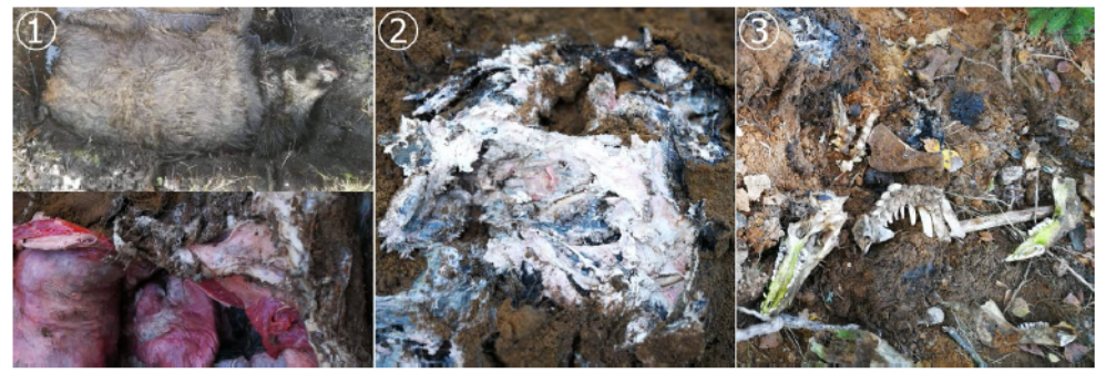
Wild boar carcass excavation including decontamination measures of the excavation place and surrounding areas has been approved by the Lithuanian Ministry of Environment and the State Food and Veterinary Service of the Republic of Lithuania. Wild boar carcasses, initially found and tested positive for ASFV infection by quantitative polymerase chain reaction (qPCR) as part of disease control measures, have been excavated during 8–11 October 2018 on 20 different locations across Lithuania (see Figure S1). Bone marrow and, if available, residual organ matrix were taken from each excavated carcass. In addition, soil was sampled on each excavation site by pooling three samples randomly taken next to the decomposing bodies. For better comparison, the decomposition status of each sampled carcass was scored. Score ① denotes that the cadaver was generally intact with closed skin and identifiable organs. Carcasses that were dismembered or open but had residual tissue obtained score ② and if only bones were left, the carcasses received score ③ (Figure 1). After excavating and sampling, the carcasses were buried again on the spot. Local soil surfaces and all involved instruments were treated using a broad-spectrum disinfectant containing peroxides, surfactants, organic acids and an inorganic buffer system (Virkon® S, Lanxess). To avoid unintentional virus inactivation of carcass samples, disinfectants were strictly applied after completion of the sampling process. The obtained specimens were subsequently tested by qPCR and virus isolation at the National Food and Veterinary Risk Assessment Institute, harbouring the National Reference Laboratory (NRL) for ASF in Lithuania and at the Friedrich-Loeffler-Institut (FLI), Germany.
 
1:尸体分解状态评分图例。①分:皮肤完好;②分:有残余组织;③分:只剩骨头。
Figure 1: Exemplary pictures of applied carcass condition score. Score ① skin closed, organs intact; Score ② residual tissue; Score  only bones.


2.2 | 实验室检测 Laboratory investigations
2.2.1 样本处理 Sample processing
采样后,样品储存在- 80°C,直到进一步使用。为了进行定量PCR和病毒分离,使用TissueLyser II (Qiagen)直径为5mm不锈钢珠在1ml磷酸盐缓冲盐水(PBS)把骨髓搅拌均匀。组织样品用杵臼在无菌砂中研磨均匀。
After collection, samples were stored at −80°C until further use. For qPCR and virus isolation, bone marrow samples were homogenized with a 5 mm stainless steel bead in 1 ml phosphate-buffered saline (PBS) using TissueLyser II (Qiagen). Tissue samples were homogenized by grinding in sterile sand with pestle and mortar.
 
2.2.2 基因组检测 Genome detection
在立陶宛国家参考实验室,使用QIAamp®RNA病毒迷你试剂盒(Qiagen)提取ASF病毒核酸。所有样本均按照King等人(2003)的方法,通过定量PCR筛选ASFV基因组。在德国弗里德里希·吕弗勒研究所,使用DNeasy®PowerSoil®Kit (Qiagen)提取了土壤和衰变基质样品的核酸。beta-actin作为内参基因,通过定量PCR (Tignon et al., 2011)对获得的洗液进行ASFV基因组筛选。此外,对土壤样本进行了猪细胞色素B基因筛选(Forth, 2015),以检查野猪DNA的存在。
At the Lithuanian NRL, ASF viral nucleic acid was extracted using the QIAamp® RNA Viral Mini Kit (Qiagen). All samples were screened for ASFV genome by qPCR according the protocol of King et al. (2003). At FLI, nucleic acids of soil and decay matrix samples have been extracted by using the DNeasy® PowerSoil® Kit (Qiagen). The obtained eluates were screened for ASFV genome by qPCR (Tignon et al., 2011) with beta-actin gene serving as internal control. In addition, soil samples were screened for swine cytochrome B gene (Forth, 2015) to check for the presence of wild boar DNA.
 
2.2.3 | 病毒分离 Virus isolation
为了检测传染性ASFV我们通过直接加入均先进行三次盲传的方式将骨髓和组织样本均匀混合,检测外周血单核(PBMC)巨噬细胞对血液的吸附。
To detect infectious ASFV, homogenized bone marrow and tissue samples were tested for hemadsorption on peripheral blood mononuclear (PBMC)-derived macrophages by directly adding the homogenate but also by running three blind passages first.
 
从健康的家猪供体猪身上采集血液,用于制备外周血单核巨噬细胞。简而言之,用Pancoll动物密度梯度培养基(PAN Biotech)edta抗凝血中获得外周血单核巨噬细胞。在37、含5% CO2的湿化环境中,用含有4-(2-羟乙基)-1-哌嗪乙磺酸(HEPES)10%胎牛血清(FCS)RPMI - 1640细胞培养基培育外周血单核巨噬细胞。培养基中添加两性霉素B、链霉素和青霉素,以避免细菌和真菌的生长。细胞培养液中加入2 ng/ml GM-CSF(粒细胞-巨噬细胞集落刺激因子Biomol)。在t12.5细胞培养瓶(Corning™)1 ml的匀浆加入外周血单核巨噬细胞(1×10⁷/ml)中进行盲传。培养48-72小时后,细胞培养瓶进行冻融,然后继续传代和检测。
Blood for the preparation of PBMC-derived macrophages was collected from healthy domestic donor pigs. In brief, PBMCs were obtained from EDTA-anti-coagulated blood using Pancoll animal density gradient medium (PAN Biotech). PBMCs were grown in RPMI-1,640 cell culture medium with 4-(2-hydroxyethyl)-1-piperazineethanesulfonic acid (HEPES) and 10% foetal calf serum (FCS) at 37°C in a humidified atmosphere containing 5% CO2. The medium was supplied with amphotericin B, streptomycin and penicillin to avoid bacterial and fungal growth. To facilitate maturation of macrophages, GM-CSF (granulocyte-macrophage colony-stimulating factor; Biomol) was added to the cell culture medium at 2 ng/ml. For blind passaging, 1 ml of the homogenate was added to PBMCderived macrophages (1 × 10⁷ cells/ml) in a T 12.5 cell culture flask (Corning™). After an incubation time of 48–72 hr, the cell culture flasks were freeze-thawed before further passaging and testing.
 
血吸附试验(HAT)按照稍作修改的标准程序进行(Carrascosa, Bustos, & Leon, 2011)简单来说,100μl分离的外周血单核巨噬细胞5×10⁶细胞/毫升的浓度被接种到96孔微孔板中(Corning™ Primaria™)16 - 24小时后,非贴壁细胞被移除,并且以上述标准补充含有GM-CSF的细胞培养液。培养24小时后,巨噬细胞开始成熟。随后,将30μl上清液的均浆样本添加到每个每个孔中加入100ul盲传的细胞培养上清液,每个样品重复测八个样。经过24小时的培育后,将20μl同源1%红细胞悬液添加到每个孔中。在3天的时间内,检测培养物对血液的吸附。当直接使用骨髓或组织匀浆时,细胞在37下经过2小时的吸附时间后,用微温的PBS洗涤细胞,而细胞培养上清液保留在细胞上,直到最后评估试验结果。以ASFV亚美尼亚2007病毒作为阳性对照。可疑结果通过细胞上清液的附加传代和定量PCR进行确认(King et al., 2003)得到证实。
The hemadsorption test (HAT) was carried out according to slightly modified standard procedures (Carrascosa, Bustos, & Leon, 2011). In brief, 100 μl of isolated PBMCs per well was seeded into a 96-well microplate (Corning™ Primaria™) at a density of 5 × 10⁶ cells/ ml. After 16–24 hr, non-adherent cells were removed and cell culture medium containing GM-CSF was replenished as described above. The culture was then incubated for 24 hr to allow initial maturation of macrophages. Subsequently, 30 μl of the supernatant of the homogenized sample was added to each well. From blind passaged samples, 100 μl of the cell culture supernatant was added to each well. Samples were tested in eight replicates. After 24 hr of incubation, 20 μl of homologous 1% erythrocyte suspension was added to each well. For readout, cultures were checked for hemadsorption over a period of 3 days. When using bone marrow or tissue homogenates directly, cells were washed after 2 hr of adsorption time at 37°C using lukewarm PBS, whereas cell culture supernatant was left on the cells until the final evaluation of the test. ASFV Armenia 2007 virus stocks were used as positive control. Doubtful results were confirmed by an additional passage and qPCR testing (King et al., 2003) of cell supernatant.
 
为了排除非血吸附ASFV毒株的存在,根据Carrascosa(2011)发表的方案,间接免疫荧光染色对每个尸体的样品进行额外检测。简而言之,就是福尔马林固定的巨噬细胞与病毒蛋白p30的单克隆抗体(由英国Pirbright研究所的Linda Dixon提供)孵育,然后用市售荧光抗体山羊抗小鼠IgG (H + L)Alexa Fluor®488 (Thermo Fisher)染色。来自在德国FLI实验室的猪的血样经主管当局(Landesamt fur Landwirtschaft, Lebensmittelsicherheit und Fischerei (LALLF) Mecklenburg-Vorpommern)批准,编号为LALLF 7221.3-2-041/17。本研究未做进一步的动物实验。使用Microsoft Excel 2007 (Microsoft)SigmaPlot进行数据分析。
To exclude the presence of non-hemadsorbing ASFV strains, samples of each carcass were additionally screened by indirect immunofluorescence staining according to the protocol published by Carrascosa et al. (2011). In brief, the formalin-fixated macrophages were incubated with monoclonal antibodies against the viral protein p30 (kindly provided by Linda Dixon, Pirbright Institute, UK) and then stained with commercially available fluorescent antibodies Goat anti-mouse IgG (H + L), Alexa Fluor® 488 (Thermo Fisher). Blood donation from pigs kept for laboratory investigation at FLI, Germany was approved by the competent authority (Landesamt für Landwirtschaft, Lebensmittelsicherheit und Fischerei (LALLF) Mecklenburg-Vorpommern) under reference number LALLF 7221.3-2-041/17. No further animal experiments have been conducted within the present study. Data analysis was performed using Microsoft Excel 2007 (Microsoft) and SigmaPlot for Windows version 11.0 (Systat software, Inc.).

【版权声明】

中国养猪网旗下所有平台转载的文章均已注明来源、中国养猪网原创文章其他平台转载需注明来源且保持图文完整性、中国养猪网特别说明的文章未经允许不可转载,感谢您的支持与配合;我们所有刊登的文章仅供养猪人参考学习,不构成投资意见。若有不妥,请及时联系我们,可添加官方微信号“zgyangzhuwang”!
详情广告
中国养猪网

中国养猪网手机APP端

精华推荐

双胞胎养猪网共建

推荐阅读

关于我们 | 服务介绍 | 招聘信息 | 版权及免责声明 | 联系我们 | 网站地图
中国农业百强网站 互联网经营备案 网信认证 网络警察 报警平台 网站备案

中国养猪网增值电信业务经营许可证:B2--20110053 中国养猪网备案号: 赣B2--20110053-5 网安备案号:36010902000949

Copyright©2010-2023 https://www.zhuwang.com.cn Inc.All Rights Reserved.新海传媒版权所有